Bridging Brain and Mind
Theories in behavioural neuroscience aim to explain how the brain underpins cognition and behaviour. With new methods for measuring brain activity, our theories linking brain to mind become increasingly precise, explaining how complex mental activities arise from networks of billions of nerve cells or neurons. In some cases models are ‘computational’, with neural processes being specified mathematically so that they can be used to make new predictions about behaviour.
This approach has great potential to break new ground in understanding how brain and mind intersect. A deep understanding of how brain-based disorders impact on cognition may have far reaching implications for rehabilitation and treatment. A shared Grand Challenge across multiple Unit programmes is the establishment of detailed brain-behaviour models across the domains of neurodevelopment, hearing, attention, memory, executive control and mental health.
CBU Research that Bridges Brain and Mind
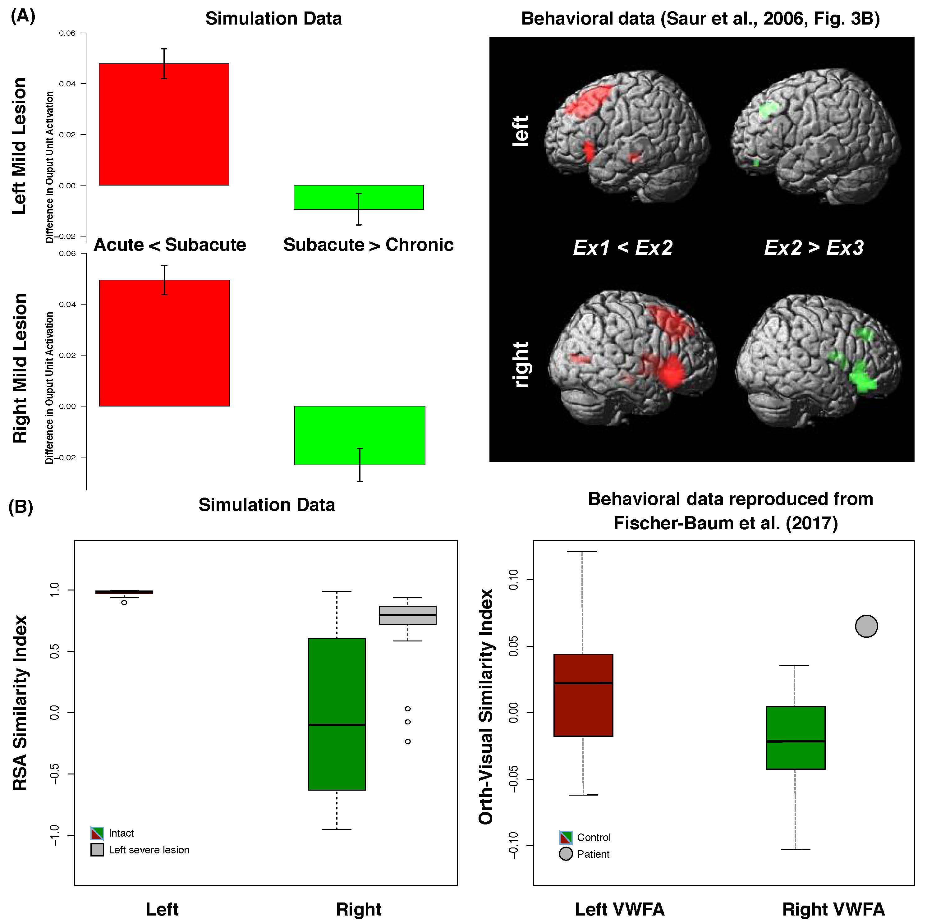
Language is a key human ability. When impaired (e.g., after stroke or neurodegeneration), patients are left with significant disability in their professional and everyday lives. These language problems (known as aphasia) are common - around one-third of the 10 million+ patients in the acute phase post stroke. Many patients show some degree of partial recovery of language in the months that follow a stroke. But what changes in the brain allow this to happen?
In this recently accepted PNAS paper, MRC CBU Scientists used a neurocomputational bilateral model of spoken language production to provide a unified explanation for a many different findings from healthy participants and in aphasia after stroke. By providing a new mechanistic account, this model provides a formal basis for understanding the variation of impairment across individual patients and also for considering options for rehabilitation
The full paper can be read here: https://www.pnas.org/content/early/2020/12/02/2010193117
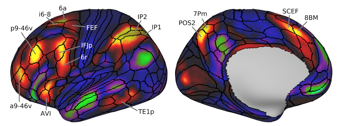
Every thought or action brings together different kinds of information. If we go to the fridge for milk, we combine knowledge of the spatial layout of the kitchen, sensory input from the contents of the fridge, our goal of making tea and our knowledge of what tea requires, as well as the commands that move our body. These different things are processed in different brain networks, and we need to sew them together into the correct sequence of operations. Using high-resolution brain imaging, our study defines a specific brain network underlying cognitive integration. With parts widely distributed across the brain, and strong connections between these parts, this network is well placed for bringing different kinds of information together. Unlike networks specialised for individual cognitive functions, such as face recognition or memory, the integration network is active during many different kinds of thought and behaviour, indicating its core role in organizing our mental lives.
Assem, M., Glasser, M. F., Van Essen, D. C., & Duncan, J. (2020). A domain-general cognitive core defined in multimodally parcellated human cortex. Cerebral Cortex, 30, 4361-4380
In the past decade, scientists have been excited about new analytical methods, using machine learning, that allow us to track information coded in patterns of brain activity. We can use them to “read” out from brain activity what participants are looking at or paying attention to, or even what they are imagining. But are the codes we read out with our machine learning algorithms actually the same codes used by the brain? To find out, CBU scientists examined how information coding changes when participants make mistakes. We found that codes in key frontoparietal brain regions predicted what sort of error a person would make, while codes elsewhere in the brain did not. This suggests a key role for activity in frontoparietal brain regions in determining behavioural performance and provides a method to determine which brain codes are meaningfully related to human behaviour.
Citation: Woolgar, A., Dermody, N., Afshar, S., Williams, M. A., & Rich, A. N. (2019). Meaningful patterns of information in the brain revealed through analysis of errors. bioRxiv, 673681.
https://www.biorxiv.org/content/10.1101/673681v1.full.pdf.
Citation: Linking the brain with behaviour: the neural dynamics of success and failure in goal-directed behaviour
Amanda K. Robinson, Anina N. Rich, Alexandra Woolgar (bioRxiv 2021)
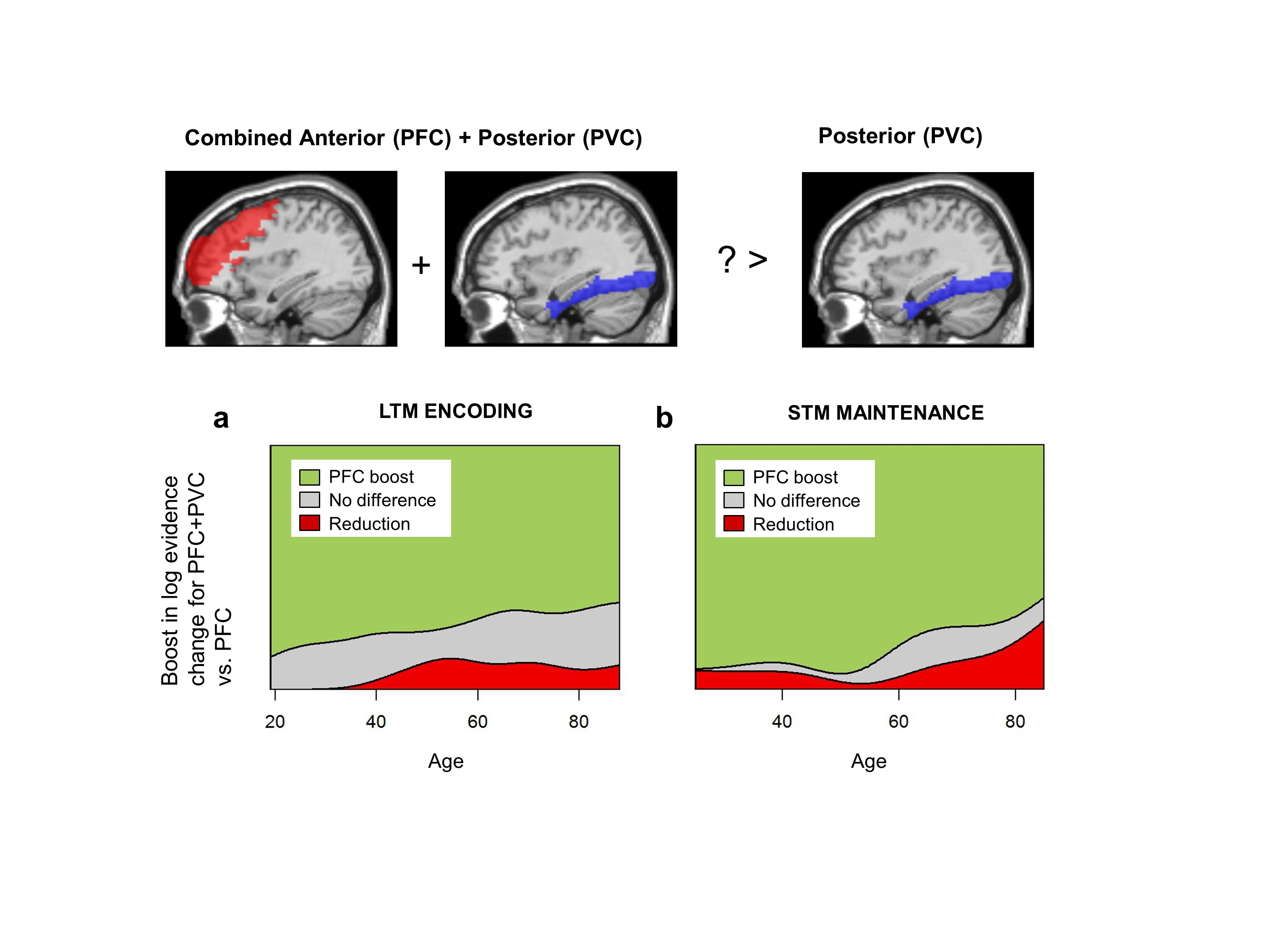
Increased prefrontal cortex activity during tasks such as encoding new memories is often observed in some healthy older adults, despite overall age-related declines in memory and other cognitive functions. This study compared two leading models of brain ageing: that the frontal cortex is either compensating for impairments elsewhere in the brain; or alternatively, that structural or neurochemical changes lead to less efficient and less specific use of resources. Using sophisticated multivariate statistical modelling of two large-scale memory datasets from Cam-CAN, the authors present evidence for the latter explanation.
Morcom, A.M., Cam-CAN & Henson, R.N. (2018). Increased prefrontal activity with aging reflects nonspecific neural responses rather than compensation. Journal of Neuroscience, 38, 7303–7313.
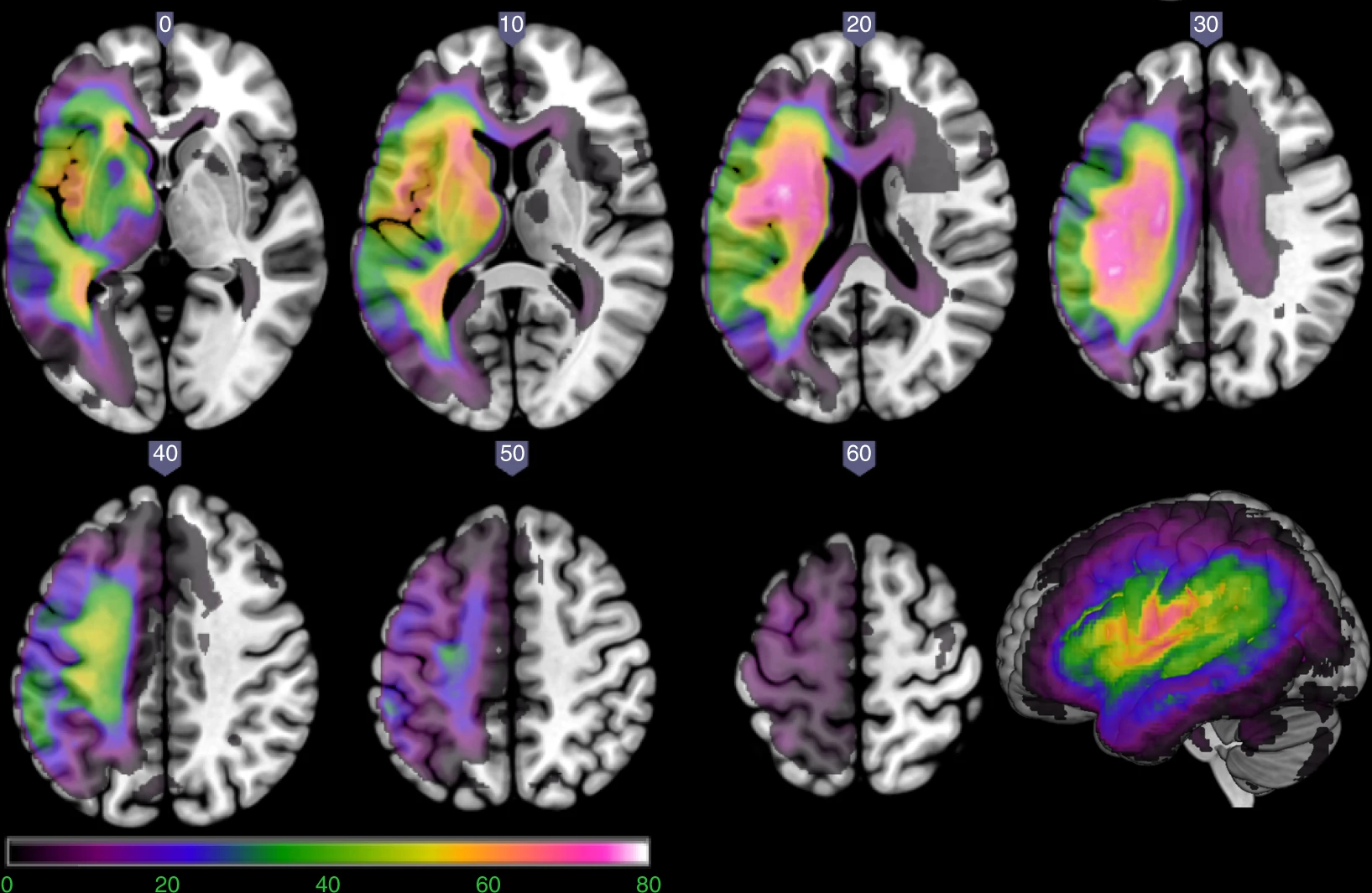
In the search for the neural basis of behaviour, researchers tend to work backwards: they first collect data about a given characteristic, then search for brain imaging metrics that correlate with that measure. But recent developments in applied mathematics and neuroimaging have enabled researchers to adopt the opposite approach. By evaluating the importance of neural features in explaining behaviour, researchers can build predictive models that get closer to establishing a causal relationship between these variables. This approach is especially powerful in cases of illnesses like stroke, when the damage to a patient’s brain occurs long before clinicians understand how it may have impaired her ability to speak and think. In this study, a team from the MRC CBU built models that predict the language abilities of patients with post-stroke aphasia. By varying three critical parameters – namely, the brain partitions, the form of neuroimaging, and the machine learning algorithm – the team was able to identify which models best predict different domains of symptoms. This study built a foundation for future work in predictive modelling, which is a critical step toward better medical care for patients with brain injuries.

Behavioural disinhibition is a common feature of frontotemporal lobar degeneration (FTLD) syndromes. Impulsive behaviour in several neuropsychiatric diseases and FTLD have been linked with reduced levels of the neurotransmitters, glutamate and GABA. This study aimed to investigate, through a transdiagnostic approach, whether prefrontal glutamate and GABA levels are reduced by FTLD and if these deficits are associated with impaired response inhibition. Participants (33 patients with FTLD syndromes, 20 control participants) completed an ultra-high field (7 T) magnetic resonance spectroscopy and a response inhibition task. Glutamate and GABA levels were measured using semi-LASER magnetic resonance spectroscopy in the right inferior frontal gyrus (IFG; response inhibition region) and the primary visual cortex (control region). FTLD patients had impaired response inhibition as well as reduced GABA concentration in the right IFG compared with controls. Glutamate concentrations did not differ between groups. Results also indicated that greater impulsivity was associated with reduced glutamate and GABA concentrations in the IFG. Glutamatergic and GABAergic deficits in the frontal lobe may be potential targets for symptomatic drug treatments for FTLD syndromes.
Murley, A. G., Rouse, M. A., Jones, P. S., Ye, R., Hezemans, F. H., O’callaghan, C., Frangou, P., Kourtzi, Z., Rua, C., Carpenter, T. A., Rodgers, C. T., & Rowe, J. B. (2020). GABA and glutamate deficits from frontotemporal lobar degeneration are associated with disinhibition. BRAIN, 143, 3449–3462. https://doi.org/10.1093/brain/awaa305
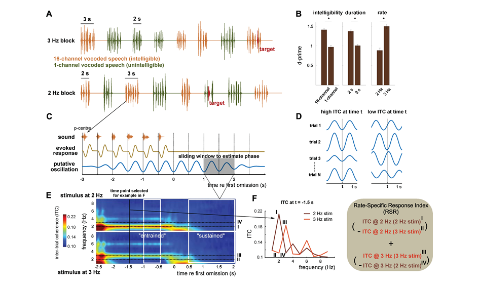
When the brain encounters a rhythm—for instance, through music, or direct electrical stimulation—it aligns its oscillation patterns (‘brainwaves’) to the beat. However, it is unclear if these stimulus-aligned brain responses are internally driven—through the brain’s own underlying patterns of activity—or if they are evoked by the rhythm of the stimulus itself. In this study, researchers aimed to clarify the difference between evoked rhythmic activity and the brain’s intrinsic oscillation patterns. Using a functional brain-imaging technique called magnetoencephalography (MEG), van Bree and colleagues looked at how brain activity changed before and after rhythmic stimulation, and whether these effects lasted longer than a rhythmic stimulus was presented. In the first experiment of this study, participants listened to intelligible and unintelligible speech. Rhythmic, intelligible speech was found to produce an oscillatory response that outlasts the stimulus in the parietal lobe of the brain. In their second experiment, the researchers stimulated participants’ brain activity using transcranial alternating current stimulation (tACS). They found that electrical stimulation changed the way that participants perceived speech: rhythmic stimulation produced rhythmic changes in speech perception. These results revealed that the effects of rhythmic brain stimulation—whether through auditory perception, or an electrical current—last beyond the duration of a stimulus, meaning that endogenous oscillations in the brain ‘carry’ the rhythms that we encounter. Therefore, endogenous neural oscillations are a key underlying feature of speech perception, and they are dependent on the rhythmic variations in speech that we encounter on a day-to-day basis. The more we know about how the brain processes speech, the closer we get to being able to treat disorders of speech perception, which affect communication and quality of life.
Citation: van Bree, S., Sohoglu, E., Davis, M.H., Zoefel, B. (2021) Sustained neural rhythms reveal endogenous oscillations supporting speech perception. PLoS-Biology, 19(2), e3001142 [Joint senior author]. https://journals.plos.org/plosbiology/article?id=10.1371/journal.pbio.3001142
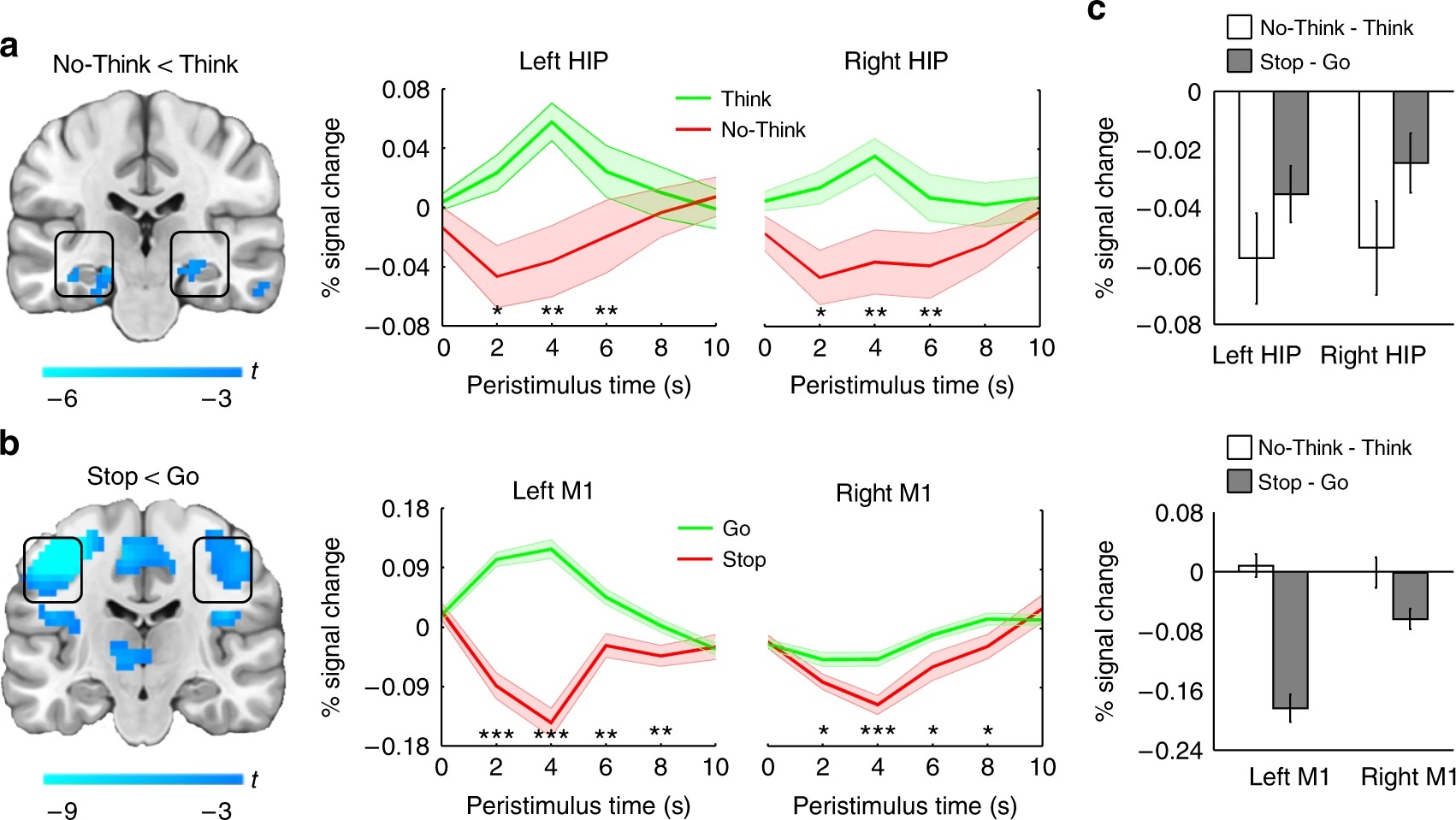
We can’t always control what we think, for those with psychiatric disorders, this is especially challenging. Intrusive memories, flashbacks, and hallucinations are hallmark symptoms of a variety of mental health conditions. Although these symptoms are often attributed to problems with brain regions that help us inhibit unwanted thoughts—such as the prefrontal cortex—difficulties in controlling intrusive thoughts are also associated with chemical differences in the brain. Hippocampal hyperactivity arising from dysfunctions in neurons that relay GABA, an inhibitory neurotransmitter, have been linked to intrusive thought patterns. In this study, researchers aimed to clarify how hippocampal GABA contributes to stopping unwanted thoughts. Participants were shown reminders of unwanted thoughts, and then tried to suppress them, while being scanned using functional MRI. The act of thought suppression led to reduced hippocampal activity. Additionally, magnetic resonance spectroscopy revealed that greater resting concentrations of hippocampal GABA predicted better control over memories. The researchers concluded that GABA activity in the hippocampus is key in the inhibition of memories, and that interneurons within the hippocampus allow us to suppress intrusive thoughts. Since intrusive thoughts are implicated within a range of psychiatric disorders, it is important that researchers and clinicians understand their neural underpinnings. In the future, this may allow for the development of more effective pharmacological and psychological treatments for negative thought patterns that are difficult to control.
Citation: Schmitz, T.W., Correia, M.M., Ferreira, C.S., Prescot, A.P., & Anderson, M.C. (2017). Hippocampal GABA enables inhibitory control over unwanted thoughts. Nature Communications, 8, 1311. doi: 10.1038/s41467-017-00956-z


 MRC Cognition and Brain Sciences Unit


