 MRC Cognition and Brain Sciences Unit
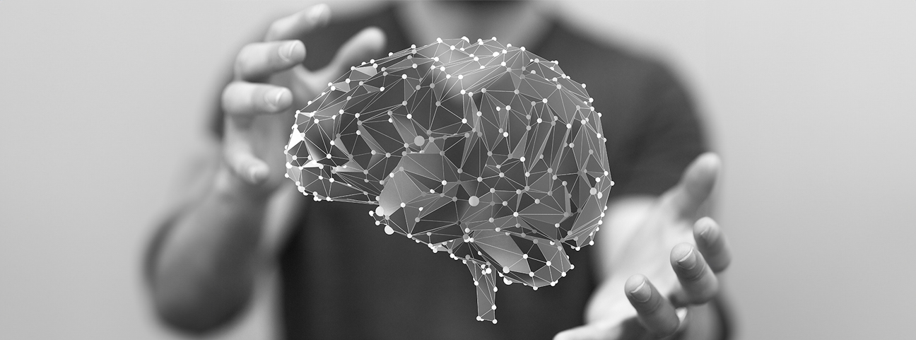


Welcome to the Cognition and Brain Sciences facilities page. Learn about our state of the art research facilities and the unique skills, knowledge and support, offered by our scientific and support teams, to both internal and external users. If you would like to know more, or have an external enquiry about using one of our facilities for clinical and cognitive neuroscience research, please click the enquire now button.
The MRC CBU MRI Facility, built in 2005 initially accommodated the first 3T Siemens Tim Trio in the UK.
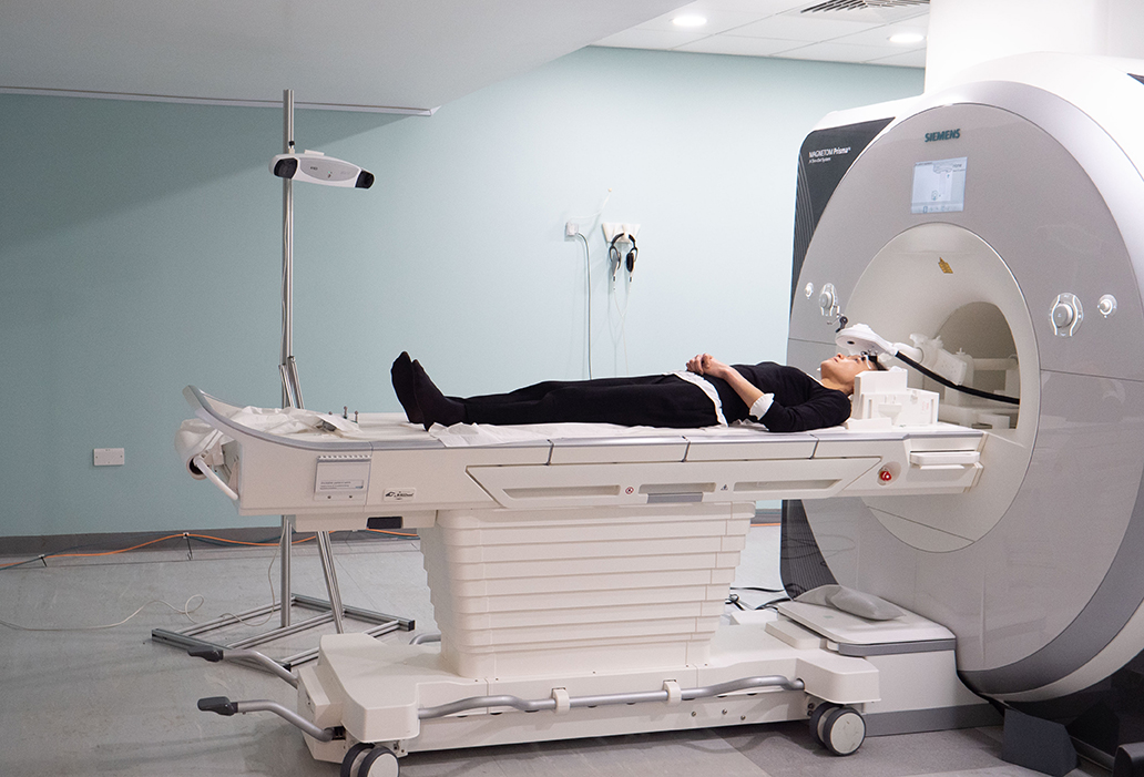
In December 2014 the trio was upgraded to a 3T Siemens Prisma, with 32- and 64-channel head coils
Situated at the end of a quiet residential street, our fMRI scanner fills a crucial role in scanning patients and healthy volunteers in an accessible and welcoming, non-medical, setting.
Our MRI scanner is widely used by research groups at the MRC CBU, across the University of Cambridge, and beyond. We also have equipment for simultaneous MRI and Transcranial Magnetic Stimulation (TMS) as well as MRI and Transcranial Ultrasound Stimulation (TUS), a setup which is unique in Cambridge and one of only a few in the world.
We have equipment for simultaneous MRI and Transcranial Magnetic Stimulation (TMS) a setup which is unique in Cambridge and one of only a few in the world. We also have equipment for Transcranial Ultrasound Stimulation (TUS), eye tracking, pulse and oxygen monitoring, and concurrent EEG. We also have a separate decommissioned scanner which can be used to help familiarise patients and volunteers to the MRI environment.
The MRI facility has a dedicated, experienced team of radiographers, methods scientists, technical and IT support and an administrator.
In the 20 years of operations the scanner facility has undertaken over 35,000 scans for over 420 individual mri studies, many of which have used the latest MRI Sequences and cutting edge mri technologies, such as TMS stimulation, EEG in mri, specialist Audio equipment and bespoke stim delivery , requiring innovative methodological approaches.
The MRI unit facilitates scanning for clinical and cognitive neuroscience research across the University of Cambridge and for external researchers including from the University of East Anglia, the University College London, Kings College London and limited commercial enterprises. We welcome scanning of healthy volunteers as well as special populations including children and patients living in the community.


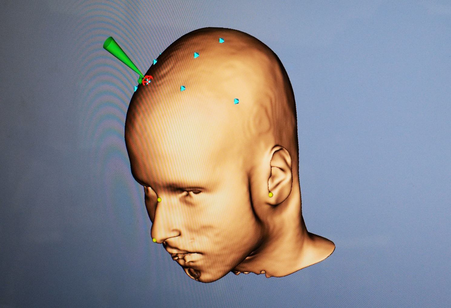
3d image of the brain from an MRI scan.
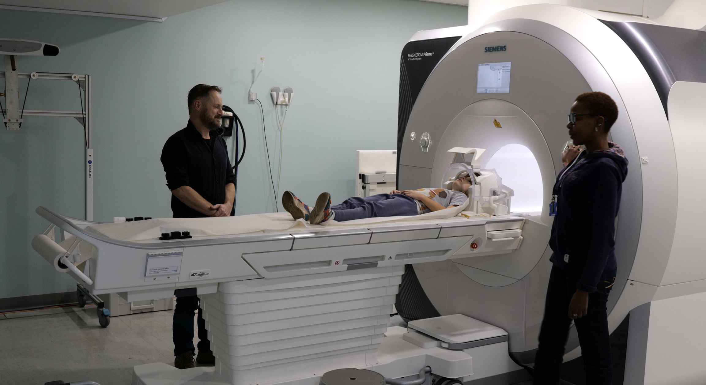
We also have a decommissioned scanner within the MRI facility, called a Mock scanner. It is used for volunteer training, technical piloting, and to help desensitise claustrophobic adults and children.
The MRC CBU Magnetoencephalograph (MEG) is one of only about ten systems of its kind in the UK. It features 306 superconducting sensors that detect minute magnetic signals generated by brain activity, offering exceptional temporal resolution at the millisecond level.
Our system enables simultaneous MEG and EEG recordings, enhancing sensitivity and spatial accuracy. It also integrates eye-tracking capabilities and can accommodate custom equipment to meet specific research needs. In 2020, we upgraded the system with a Helium recycling facility, reducing costs and improving environmental sustainability.
The MEG suite is designed to provide a quiet and calming environment. Conveniently located on a residential street, it is easily accessible for adult participants of all ages, as well as for patients and special populations from the community.
A skilled team of operators and support staff maintains the system, offering comprehensive training for new researchers. This includes guidance on everything from stimulus presentation and data collection to advanced data analysis.
Over the past 18 years, we have conducted more than 6,500 recordings for over 150 MEG studies. These projects involve CBU researchers, university departments, visiting scientists, and industry collaborators. Our research spans a diverse range of topics, including cognition, language, dementia, schizophrenia, and developmental and aging studies.

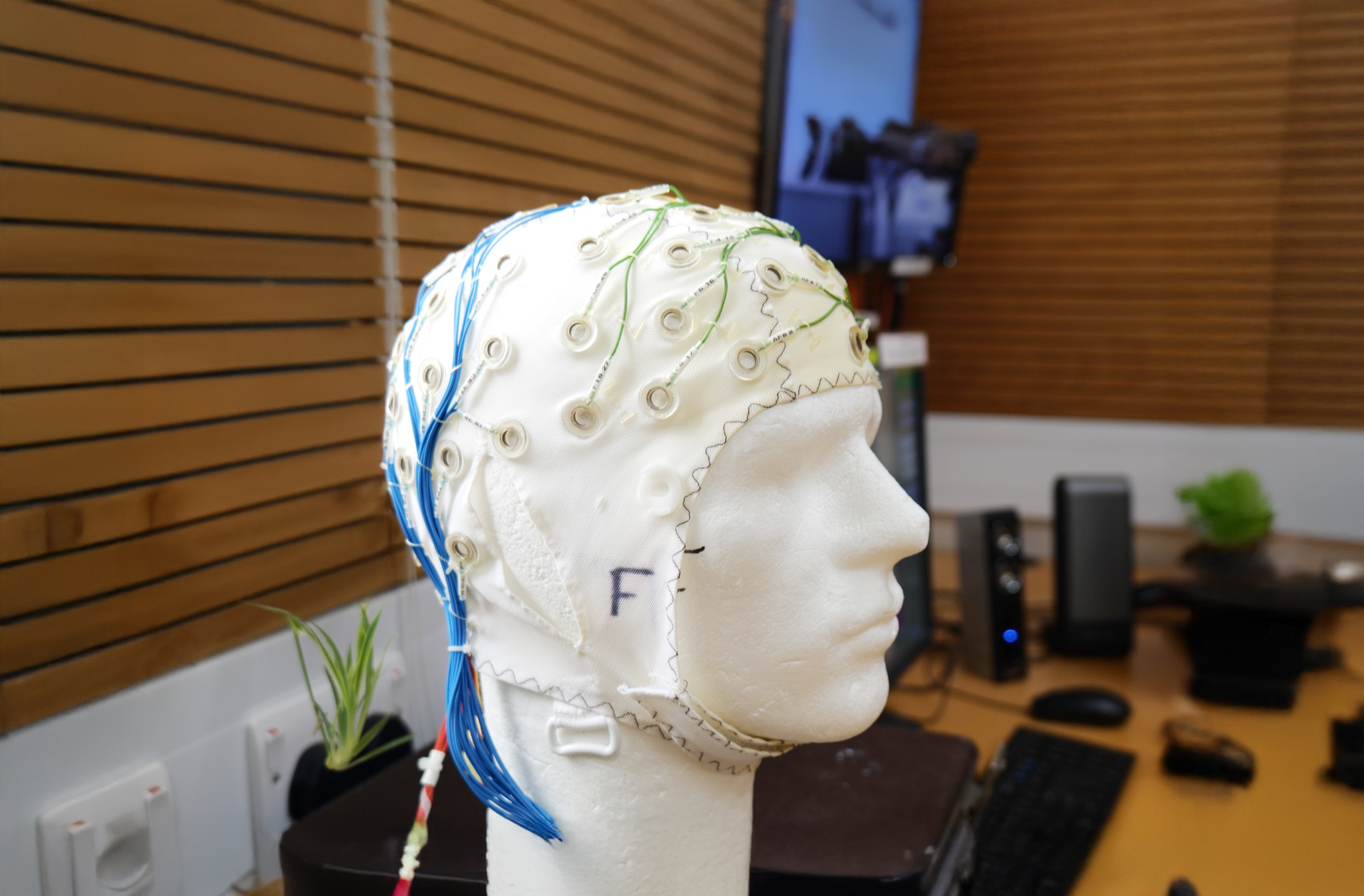
Cap used by participants for EEG and MEG experiemnts.
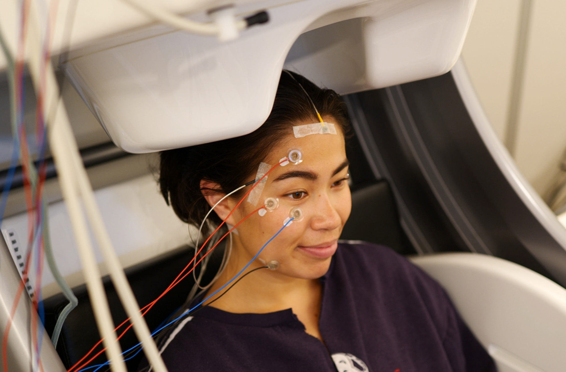
Participant in the MEG machine ready to be scanned.
Our state-of-art TMS facility is the newest addition to the CBU and is unique in the extent of its capabilities. A recent grant award facilitated the purchase of a robotic aid, which had improved both research efficiency and the participant experience. In addition to stand-alone TMS, our TMS lab can support concurrent neural measurement with EEG, complementing the MR-TMS. The CBU is actively involved in working with emerging neurostimulation technologies and so we have developed this facility with the future in mind – ready to meet the next generation of challenges in cognitive science.

TMS plotting
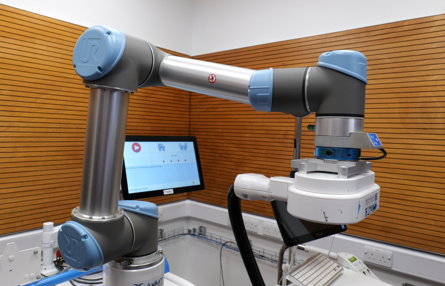
TMS robotic aid
MRI with TMS
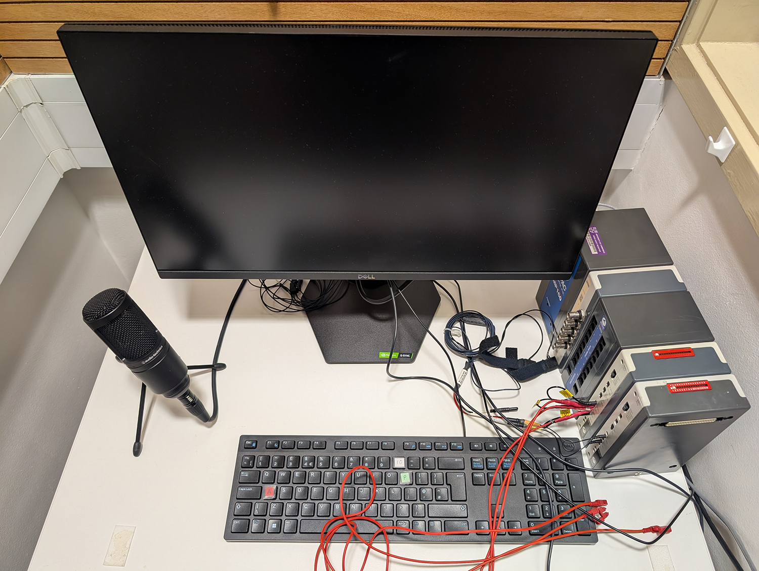
The sound laboratory features a wide range of equipment for performing auditory experiments, including three IAC double-walled sound insulating booths, each connected to a dedicated test rig. Stimulus calibration devices include a LeCroy Waverunner 4-channel digital storage oscilloscope, an HP3561A dynamic signal analyzer, a B&K 4153 artificial ear, and a KEMAR mannikin that can be used for calibrating stimuli presented either in free field or through headphones. Research software and hardware for presenting arbitrary electrical stimuli via each of the main three models of cochlear implant are also available.
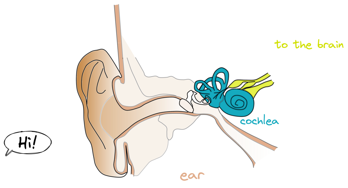
Diagram of how a cochlear implant works.
The MRC CBU currently runs two psychophysiology laboratories, using BIOPAC MP100 systems to record heart rate (HR), galvanic skin response (GSR), and electromyography (EMG) data. These serve as valuable measures of peripheral nervous system function in a variety of experimental designs. We also have a BIOPAC MP150 system that can measure GSR and HR in the fMRI environment. Data is analysed using BIOPAC Acqknowledge software and also via software developed in-house.
The MRC Cognition and Brain Sciences Unit (CBU) is equipped with three high-speed eye trackers (SR Research EyeLink systems), supporting a wide range of research applications. Two eye-trackers are customised for use in our EEG/MEG and fMRI labs, respectively, and one is available for behavioural experiments. Each system supports sampling rates of up to 1000 Hz, offering high temporal resolution suitable for precise eye movement research, e.g. using reading or visual search paradigms. These eye trackers are routinely used by multiple research groups and are compatible with a variety of stimulus presentation and data analysis software packages, ensuring flexibility across experimental paradigms.
The MRC CBU hosts several wiki pages offering a wealth of technical and methodological information relating to collection and analysis of neuroimaging data. Use one of the images below to visit a specific wiki, or go to the main wiki farm located here.


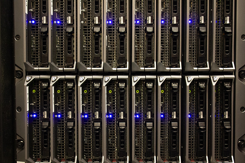
The MRC CBU uses computers in nearly all aspects of our research activities. This ranges from data collection using stimulus delivery computers for testing volunteers to control systems for our fMRI and MEG brain scanners and other laboratory equipment. Once collected the data is sent to be stored in our large scale data storage system ready for analysis on our HPC (High Performance Compute) cluster. Then scientists analyse this data and write papers from numerous desktop computers or even on a laptop the other side of the world while preparing to give a talk. The computing group build and maintain all these services while ensuring staff are able to use these services and resources effectively.
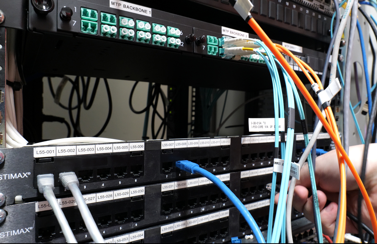
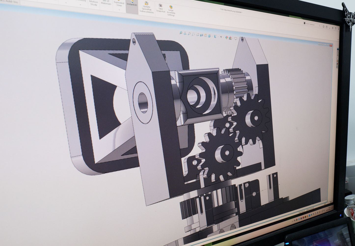
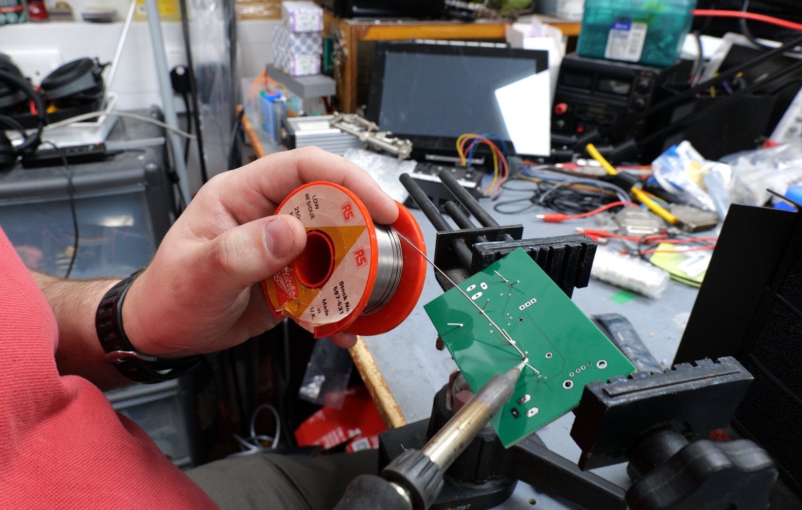
The technical department provide support for staff in designing, prototyping, building, repairing equipment/software for use in experiments or where else needed. This could be from a simple button box to a more complicated eye tracking experiment. They also manage item procurement, the processing of goods coming in and out, health and safety and making sure the equipment, building and you are as safe as possible.
Also part of this team is the Graphics officer who is in charge of looking after all areas of media such as AV, video, illustration, posters, web, photography and 3D modelling.